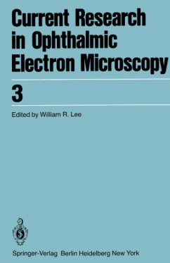
Current Research in Ophthalmic Electron Microscopy
Herausgegeben von Lee, W. B.
Current Research in Ophthalmic Electron Microscopy
Herausgegeben von Lee, W. B.
- Broschiertes Buch
- Merkliste
- Auf die Merkliste
- Bewerten Bewerten
- Teilen
- Produkt teilen
- Produkterinnerung
- Produkterinnerung
Transactions of the Seventh Annual Meeting of the European Club for Ophtalmic Fine Structure in Ystad, Sweden, April 20 and 21, 1979
Andere Kunden interessierten sich auch für
![Transactions of the 8th Annual Meeting of the European Club for Ophthalmic Fine Structure in West Berlin, March 28 and 29,1980 Transactions of the 8th Annual Meeting of the European Club for Ophthalmic Fine Structure in West Berlin, March 28 and 29,1980]() Transactions of the 8th Annual Meeting of the European Club for Ophthalmic Fine Structure in West Berlin, March 28 and 29,198077,99 €
Transactions of the 8th Annual Meeting of the European Club for Ophthalmic Fine Structure in West Berlin, March 28 and 29,198077,99 €![Electron Microscopy of the Kidney Electron Microscopy of the Kidney]() Anil K. MandalElectron Microscopy of the Kidney100,99 €
Anil K. MandalElectron Microscopy of the Kidney100,99 €![The Testicular Descent in Human The Testicular Descent in Human]() K.J. BarteczkoThe Testicular Descent in Human53,49 €
K.J. BarteczkoThe Testicular Descent in Human53,49 €![The Human Nasolacrimal Ducts The Human Nasolacrimal Ducts]() F. PaulsenThe Human Nasolacrimal Ducts77,99 €
F. PaulsenThe Human Nasolacrimal Ducts77,99 €![Diagnostic Electron Microscopy Diagnostic Electron Microscopy]() Richard G. DickersinDiagnostic Electron Microscopy77,99 €
Richard G. DickersinDiagnostic Electron Microscopy77,99 €![Atlas of the Ear Atlas of the Ear]() Yasuo HaradaAtlas of the Ear77,99 €
Yasuo HaradaAtlas of the Ear77,99 €![Membrane Physiology Membrane Physiology]() Thomas E. Andreoli / Darrell D. Fanestil / Joseph F. Hoffman / Stanley G. Schultz (eds.)Membrane Physiology62,99 €
Thomas E. Andreoli / Darrell D. Fanestil / Joseph F. Hoffman / Stanley G. Schultz (eds.)Membrane Physiology62,99 €-
-
-
Transactions of the Seventh Annual Meeting of the European Club for Ophtalmic Fine Structure in Ystad, Sweden, April 20 and 21, 1979
Produktdetails
- Produktdetails
- Current Research in Ophthalmic Electron Microscopy .3
- Verlag: Springer / Springer Berlin Heidelberg / Springer, Berlin
- Artikelnr. des Verlages: 978-3-540-09953-6
- 1980.
- Seitenzahl: 172
- Erscheinungstermin: 1. April 1980
- Englisch
- Abmessung: 244mm x 170mm x 10mm
- Gewicht: 320g
- ISBN-13: 9783540099536
- ISBN-10: 3540099530
- Artikelnr.: 36111759
- Herstellerkennzeichnung
- Springer-Verlag KG
- Sachsenplatz 4-6
- 1201 Wien, AT
- ProductSafety@springernature.com
The Development of the Irido-corneal Angle in the Chick Embryo.- Immunoelectronmicroscopical Investigations on Isolated Collagen Fibrils.- Combined Macular Dystrophy and Cornea Guttata: An Electron Microscopic Study.- Age Related Changes in Extracellular Materials in the Inner Wall of Schlemm's Canal.- Preliminary Observations on Human Trabecular Meshwork Cells in vitro.- Transcellular Aqueous Humor Outflow: A Theoretical and Experimental Study.- Increased Vascular Permeability in the Rabbit Iris Induced by Prostaglandin E1. An Electron Microscopic Study Using Lanthanum as a Tracer in vivo.- Frozen Resin-Cracking, Dry-Cracking and Enzyme-Digestion Methods in SEM as Applied to Ocular Tissues.- Scanning Electron Microscopy of Frozen-Cracked, Dry-Cracked and Enzyme-Digested Tissue of Human Malignant Choroidal Melanomas.- Vitreous Membrane Formation After Experimental Vitreous Haemorrhage.- Cellular Decay in the Rat Retina During Normal Post-natal Development: A Preliminary Quantitative Analysis of the Basic Endogenous Rhythm.- Scanning Electron Microscopy of Frozen-Cracked, Dry-Cracked, and Enzyme-Digested Retinal Tissue of a Monkey (Cercopithecus Aethiops) and of Man.- Recovery of the Rabbit Retina After Light Damage (Preliminary Observations).- The Retina in Lafora Disease: Light and Electron Microscopy.- Indexed in Current Contents.
The Development of the Irido-corneal Angle in the Chick Embryo.- Immunoelectronmicroscopical Investigations on Isolated Collagen Fibrils.- Combined Macular Dystrophy and Cornea Guttata: An Electron Microscopic Study.- Age Related Changes in Extracellular Materials in the Inner Wall of Schlemm's Canal.- Preliminary Observations on Human Trabecular Meshwork Cells in vitro.- Transcellular Aqueous Humor Outflow: A Theoretical and Experimental Study.- Increased Vascular Permeability in the Rabbit Iris Induced by Prostaglandin E1. An Electron Microscopic Study Using Lanthanum as a Tracer in vivo.- Frozen Resin-Cracking, Dry-Cracking and Enzyme-Digestion Methods in SEM as Applied to Ocular Tissues.- Scanning Electron Microscopy of Frozen-Cracked, Dry-Cracked and Enzyme-Digested Tissue of Human Malignant Choroidal Melanomas.- Vitreous Membrane Formation After Experimental Vitreous Haemorrhage.- Cellular Decay in the Rat Retina During Normal Post-natal Development: A Preliminary Quantitative Analysis of the Basic Endogenous Rhythm.- Scanning Electron Microscopy of Frozen-Cracked, Dry-Cracked, and Enzyme-Digested Retinal Tissue of a Monkey (Cercopithecus Aethiops) and of Man.- Recovery of the Rabbit Retina After Light Damage (Preliminary Observations).- The Retina in Lafora Disease: Light and Electron Microscopy.- Indexed in Current Contents.

