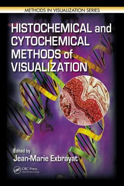
The book begins by discussing techniques in light microscopy. It reviews classical methods of visualization, histochemical and histoenzymatic methods, and methods used to visualize cell proliferation and apoptosis. Next, the book examines the cytochemical methods used in electron microscopy with traditional techniques, as well as more specialized methods. The final section provides an overview of image analysis and describes how image processing methods can be used to extract vital information. A 16-page insert supplies color illustrations to enhance the text.
Techniques will continue to adapt to the latest technological innovations, allowing more and more precise quantification of images. These developments are essential to the biological as well as the medical sciences. This manual is a critical resource for novice and experienced researchers, technicians, and students who need to visualize what happens in the cell, the molecules expressed, the main enzymatic activities, and the repercussions of the molecular activities upon the structure of the cells in the body.
Dieser Download kann aus rechtlichen Gründen nur mit Rechnungsadresse in A, B, BG, CY, CZ, D, DK, EW, E, FIN, F, GR, HR, H, IRL, I, LT, L, LR, M, NL, PL, P, R, S, SLO, SK ausgeliefert werden.

