Adrian Kendal Dixon, David J. Bowden, Bari M. Logan, Harold Ellis
Human Sectional Anatomy (eBook, ePUB)
Pocket atlas of body sections, CT and MRI images, Fourth edition
42,95 €
42,95 €
inkl. MwSt.
Sofort per Download lieferbar
21 °P sammeln
42,95 €
Als Download kaufen
42,95 €
inkl. MwSt.
Sofort per Download lieferbar
21 °P sammeln
Jetzt verschenken
Alle Infos zum eBook verschenken
42,95 €
inkl. MwSt.
Sofort per Download lieferbar
Alle Infos zum eBook verschenken
21 °P sammeln
Adrian Kendal Dixon, David J. Bowden, Bari M. Logan, Harold Ellis
Human Sectional Anatomy (eBook, ePUB)
Pocket atlas of body sections, CT and MRI images, Fourth edition
- Format: ePub
- Merkliste
- Auf die Merkliste
- Bewerten Bewerten
- Teilen
- Produkt teilen
- Produkterinnerung
- Produkterinnerung
Bitte loggen Sie sich zunächst in Ihr Kundenkonto ein oder registrieren Sie sich bei
bücher.de, um das eBook-Abo tolino select nutzen zu können.
Hier können Sie sich einloggen
Sie sind bereits eingeloggt. Klicken Sie auf 2. tolino select Abo, um fortzufahren.
Bitte loggen Sie sich zunächst in Ihr Kundenkonto ein oder registrieren Sie sich bei bücher.de, um das eBook-Abo tolino select nutzen zu können.
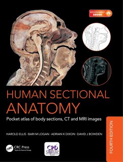
Now in its fourth edition, this portable guide and essential learning aid now contains new material. As with the previous editions, the superb full-color cadaver sections are compared with CT and MRI images. The radiological images have all been replaced with new examples for this latest edition, taken on the most up-to date equipment to ensure excellent visualization of the anatomy. The photographic material is enhanced by useful notes with details of important anatomical and radiological features.
- Geräte: eReader
- mit Kopierschutz
- eBook Hilfe
- Größe: 106.18MB
Andere Kunden interessierten sich auch für
- Alain C. MasqueletAtlas of Surgical Anatomy (eBook, ePUB)62,95 €
- David HeylingsMcMinn's Concise Human Anatomy (eBook, ePUB)26,95 €
- Kinesh PatelComplete Revision Notes for Medical and Surgical Finals (eBook, ePUB)28,95 €
- Alexander KingFirst FRCR Anatomy Examination Revision (eBook, ePUB)49,95 €
- Shaun QuigleyFirst FRCR Anatomy Practice Examinations (eBook, ePUB)49,95 €
- Rosie JonesInterpreting Musculoskeletal Images (eBook, ePUB)34,95 €
- Phillip YoongMnemonics for Radiologists and FRCR 2B Viva Preparation (eBook, ePUB)50,95 €
-
-
-
Now in its fourth edition, this portable guide and essential learning aid now contains new material. As with the previous editions, the superb full-color cadaver sections are compared with CT and MRI images. The radiological images have all been replaced with new examples for this latest edition, taken on the most up-to date equipment to ensure excellent visualization of the anatomy. The photographic material is enhanced by useful notes with details of important anatomical and radiological features.
Dieser Download kann aus rechtlichen Gründen nur mit Rechnungsadresse in A, B, BG, CY, CZ, D, DK, EW, E, FIN, F, GR, HR, H, IRL, I, LT, L, LR, M, NL, PL, P, R, S, SLO, SK ausgeliefert werden.
Produktdetails
- Produktdetails
- Verlag: Taylor & Francis eBooks
- Seitenzahl: 288
- Erscheinungstermin: 17. Oktober 2017
- Englisch
- ISBN-13: 9781498708708
- Artikelnr.: 55377942
- Verlag: Taylor & Francis eBooks
- Seitenzahl: 288
- Erscheinungstermin: 17. Oktober 2017
- Englisch
- ISBN-13: 9781498708708
- Artikelnr.: 55377942
- Herstellerkennzeichnung Die Herstellerinformationen sind derzeit nicht verfügbar.
Harold Ellis, emeritus professor of surgery, Guy's, King's and St. Thomas' School of Biomedical Sciences, London, UK
Bari M. Logan, former university prosector, Department of Anatomy, University of Cambridge; prosector, Department of Anatomy, Royal College of Surgeons of England, London; and anatomical preparator, Department of Human Morphology, University of Nottingham, UK
Adrian Dixon, professor emeritus of radiology, University of Cambridge; honorary consultant radiologist, Addenbrooke's Hospital; and Master, Peterhouse, Cambridge, UK
David J. Bowden, radiology registrar, Department of Radiology, University of Cambridge School of Clinical Medicine, Addenbrooke's Hospital, Cambridge, UK
BRAIN. Superficial dissection. Selected images. HEAD. Axial sections
(male). Selected images: axial MRI. Selected images: temporal bone/inner
ear: axial CT. Coronal sections (female). Sagittal sections (male). NECK.
Axial sections (female). Sagittal sections (male). THORAX. Axial sections
(male). Axial sections (female). Selected images: heart. Selected images:
mediastinum: axial CT. Selected images: coronal MRI. Selected images:
coronal chest CT. Selected images: coeliac and great vessels. ABDOMEN.
Axial sections (male). Axial sections (female). Selected images: lumbar
spine: axial CT. Selected images: lumbar spine: coronal MRI. Selected
images: lumbar spine: sagittal MRI. PELVIS. Axial sections (male). Selected
images: coronal MRI (male). Axial sections (female). Selected images: axial
MRI (female). Selected images: coronal MRI (female). Selected images:
sagittal MRI (female). Selected images: colon. Selected images: coronal
abdominal CT. LOWER LIMB. Hip: coronal sections (female). Selected images:
pelvic girdle. Thigh: axial sections (male). Knee: axial sections (male).
Knee: coronal sections (male). Knee: sagittal sections (female). Leg: axial
sections (male). Ankle: axial sections (male). Ankle: coronal sections
(female). Foot: sagittal sections (male). Foot: coronal sections (male).
UPPER LIMB. Shoulder: axial sections (female). Shoulder: coronal sections
(male). Arm: axial sections (male). Elbow: axial sections (male). Elbow:
coronal sections (female). Forearm: axial sections (male). Wrist: axial
sections (male). Hand: coronal sections (female). Hand: sagittal sections
(female). Hand: axial sections (male). Selected images: shoulder girdle.