73,95 €
73,95 €
inkl. MwSt.
Sofort per Download lieferbar
payback
37 °P sammeln
73,95 €
73,95 €
inkl. MwSt.
Sofort per Download lieferbar

Alle Infos zum eBook verschenken
payback
37 °P sammeln
Als Download kaufen
73,95 €
inkl. MwSt.
Sofort per Download lieferbar
payback
37 °P sammeln
Jetzt verschenken
73,95 €
inkl. MwSt.
Sofort per Download lieferbar

Alle Infos zum eBook verschenken
payback
37 °P sammeln
  • Format: PDF

This book presents MRI pulse sequences using a simple and easy to understand approach. The use of magnetic resonance imaging (MRI) is growing exponentially, in part because of the excellent anatomic and pathologic detail provided by the modality and recent technologic advances that have led to faster acquisition times. Radiology residents are now introduced in their 1st year of training to the MR pulse sequences routinely used in clinical imaging, including various spin-echo, gradient-echo, inversion-recovery, echo-planar imaging, and MR angiographic sequences. In this book, expert authors…mehr

  • Geräte: PC
  • ohne Kopierschutz
  • eBook Hilfe
  • Größe: 78.19MB
Produktbeschreibung
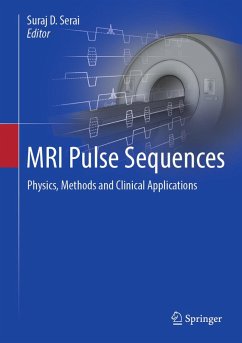
This book presents MRI pulse sequences using a simple and easy to understand approach. The use of magnetic resonance imaging (MRI) is growing exponentially, in part because of the excellent anatomic and pathologic detail provided by the modality and recent technologic advances that have led to faster acquisition times. Radiology residents are now introduced in their 1st year of training to the MR pulse sequences routinely used in clinical imaging, including various spin-echo, gradient-echo, inversion-recovery, echo-planar imaging, and MR angiographic sequences. In this book, expert authors describe the physical basis of the most common MR pulse sequences routinely used in clinical MRI. The complexity of MRI pulse sequences requires expert knowledge about the underlying physics and contrast mechanisms to select from the wide range of available applications and protocols. The pulse sequences feature high versatility concerning the generation of different tissue contrasts using different sequences of RF and magnetic field gradient events. To enhance understanding, explanation of the physical principles will be simplified, and to highlight their practical relevance, clinical examples will be interspersed throughout the book. Several roadblocks still slow the pace of progress, particularly inefficient education of MR researchers, and lack of harmonization of approaches that limits the understanding, development, and application of pulse sequences among multiple clinical and research groups. With the help of this book, these limitations are addressed by providing a comprehensive collection of chapters on MRI pulse sequence methods that will serve as a foundational resource for application of these methods on clinical MRI. This is an ideal guide for radiologists and trainees working with MRI.


Dieser Download kann aus rechtlichen Gründen nur mit Rechnungsadresse in A, B, BG, CY, CZ, D, DK, EW, E, FIN, F, GR, HR, H, IRL, I, LT, L, LR, M, NL, PL, P, R, S, SLO, SK ausgeliefert werden.

Autorenporträt
Dr. Serai is an MRI Scientist and Magnetic Resonance Safety Expert, and an Associate Professor of Radiology at the University of Pennsylvania. He received his Bachelor's Degree in Engineering from the University of Mumbai, India, and his Masters of Science and Ph.D. Degrees from the University of Illinois, Chicago. Dr. Serai is an active member of the International Society for Magnetic Resonance (ISMRM), the Radiological Society of North America (RSNA), the American Association of Physicists in Medicine (AAPM), American College of Radiology (ACR), Quantitative Imaging Bio-Markers Alliance (QIBA), and Society for Pediatric Radiology (SPR). Dr. Serai has widely published and additionally serves as an Ad Hoc Peer Reviewer for Pediatric Radiology, Radiology, American Journal of Roentgenology, Academic Radiology, Magnetic Resonance in Medicine, Abdominal Imaging, and Journal of Magnetic Resonance in Medicine. Dr. Serai's background allows him to work closely with radiologists to develop protocol, measure quantitative MRI values using mono and bi-exponential decays, and generate visual maps to demonstrate changes in regional pathophysiology, which ultimately translate to clinical application and outcome improvement. Over the last 20 years, Dr. Serai has developed and applied MR imaging-based methods for Neuro, iron and fat quantification in the body, elastography, and T1/T2 mapping techniques to suit pediatric needs.